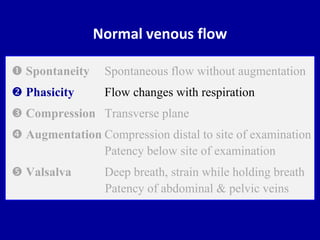
Best Describe Spontaneous and Phasic Flow in a Vein
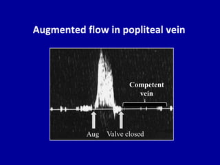
In addition special attention should be paid to the status of the. Duplex ultrasound findings in acute DVT consist of noncompressibility of the vein partial or absent color flow in the lumen visualization of luminal thrombus absence of phasic variation with respiration and lack of augmentation of venous flow with calf compression and usually dilatation of the vein.
2

Spontaneous And Phasic Venous Flow Waveform On Ultrasound Google Search Ultrasound Medical Ultrasound Diagnostic Medical Sonography

Side Difference Of The Venous Flow In The Distal Subclavian Vein In A Download Scientific Diagram
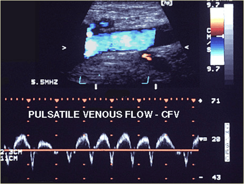
Doppler Ultrasound Of Normal Venous Flow

Doppler Interrogation Of The Femoral Vein In The Critically Ill Patient The Fastest Potential Acoustic Window To Diagnose Right Ventricular Dysfunction Abstract Europe Pmc
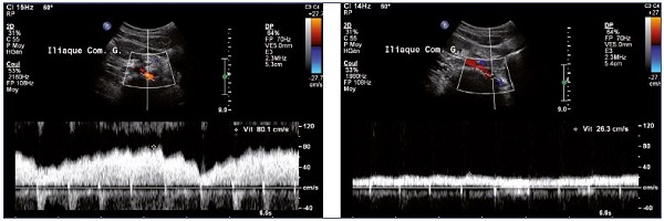
Doppler Ultrasound Of Normal Venous Flow

An Example Of Normal Respiratory Phasicity Of Venous Fl Ow As The Download Scientific Diagram
Doppler Ultrasound Of Normal Venous Flow
Role Of Duplex Ultrasound Investigation In The Management Of Postthrombotic Syndrome Servier Phlebolymphologyservier Phlebolymphology

Spectral Doppler Waveform Analysis Of The External Iliac Vein Eiv Download Scientific Diagram

Spectral Doppler Waveform Analysis Of The Lower Limb Veins Spontaneous Download Scientific Diagram
2

Upper Extremity Venous Evaluation Iame

The Importance Of Monophasic Doppler Waveforms In The Common Femoral Vein Lin 2007 Journal Of Ultrasound In Medicine Wiley Online Library

Colour Flow Doppler And Pulsed Doppler Spectral Waveform At The Right Download Scientific Diagram

Diagnosis Of Iliac Vein Obstruction With Duplex Ultrasound Endovascular Today

Upper Extremity Venous Evaluation Iame
Bilateral Lower Extremity Flashcards Easy Notecards

Upper Extremity Venous Evaluation Iame

Vascular Review Ultrasound Flashcards Quizlet


Comments
Post a Comment